Bacterial structure and morphology by Dr. Shireen Rafiq (RMC)
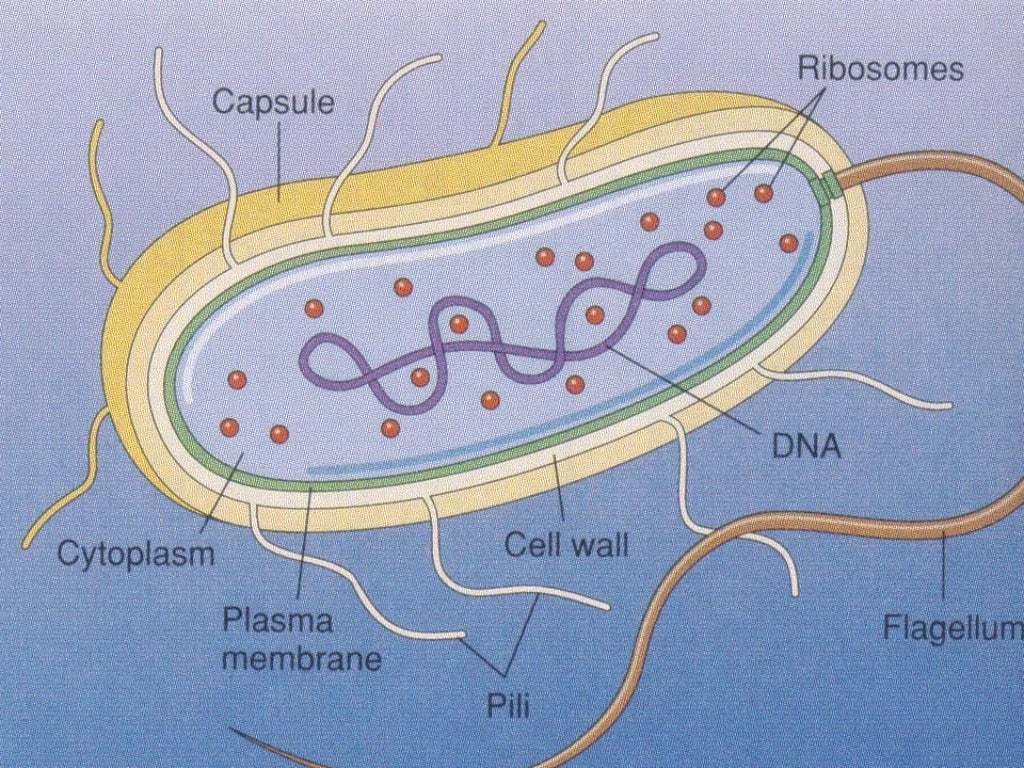
Most bacteria aren't harmful, but certain types can make you sick. 800.223.2273;. They're microbes with a very simple cell structure. Bacteria have cell walls. Within the cell walls, a bacteria diagram would show the structure of each cell. Each bacterium contains cytoplasm, ribosomes and DNA. Outside the cell wall, one or more bacteria.
Bacteria Grade 11 Biology Study Guide
These are thin, short filaments (0.1-1.5 μm x 4 to 8 nm) extruding from the cytoplasmic membrane, also called pili. They are made of protein (pilin). It is an outer covering of thin jelly-like material (0.2 μm in width) that surrounds the cell wall. Only some bacterial species possess capsule.
Cellular Structure of Bacteria ZeroInfections
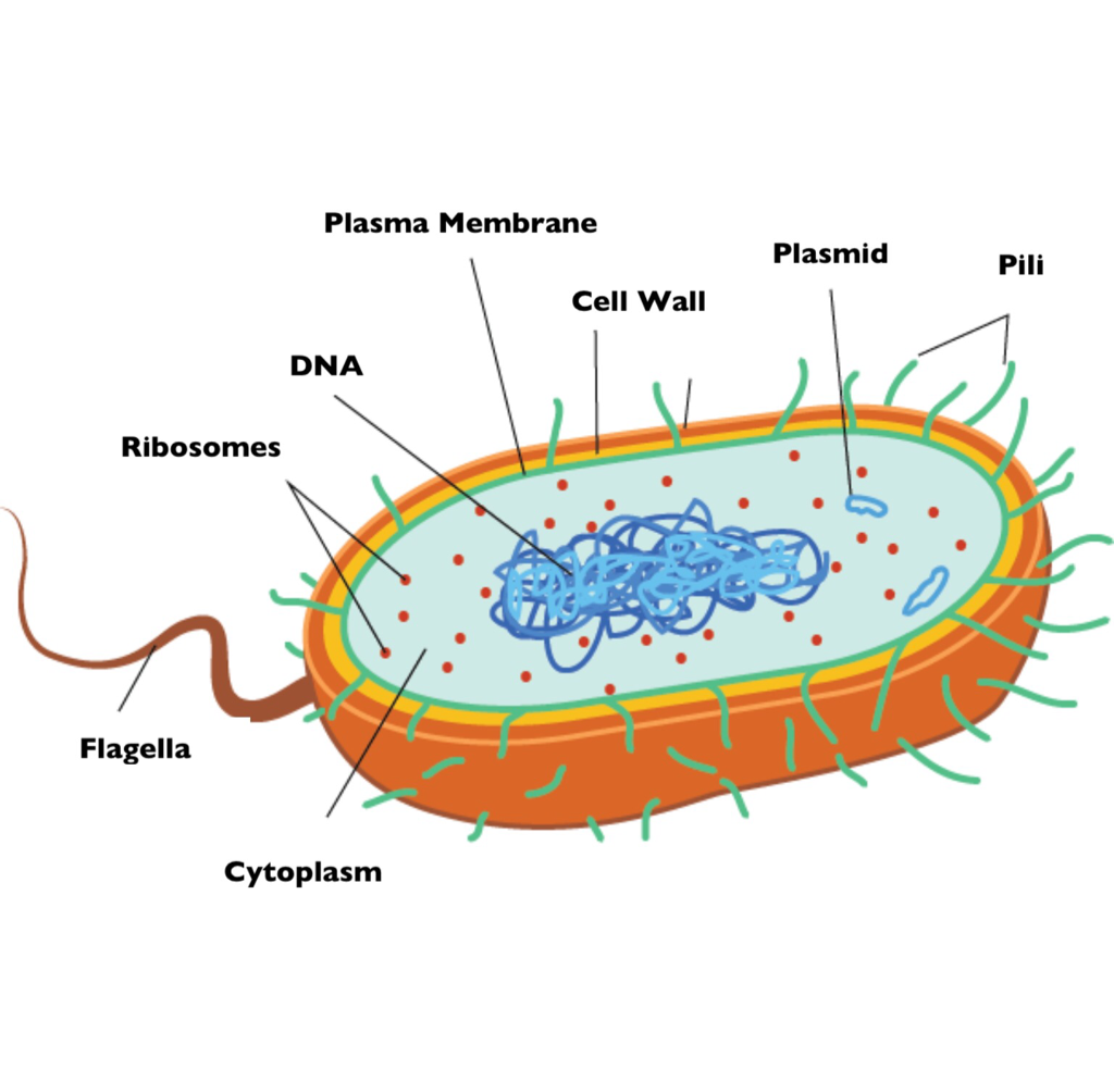
DNA in a nucleus. Plasmids are found in a few simple eukaryotic organisms. Prokaryotic cell (bacterial cell) DNA is a single molecule, found free in the cytoplasm. Additional DNA is found on one.

Bacteria Cell Structure YouTube
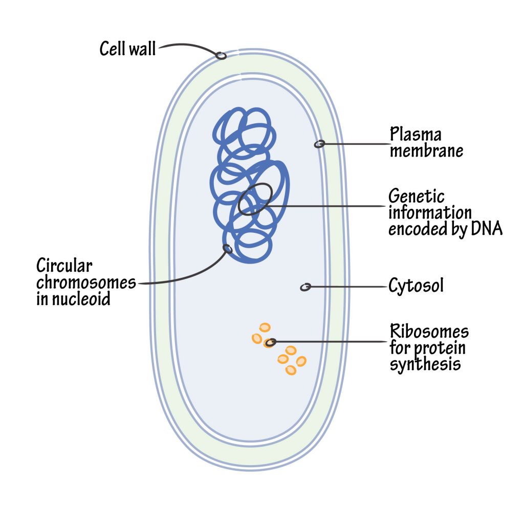
Bacteria are microscopic, unicellular, prokaryotic organisms. They do not have membrane-bound cell organelles and lack a true nucleus, hence are grouped under the domain "Prokaryota " together with Archae. In a three-domain system, Bacteria is the largest domain. ( Living beings are classified into Archae, Bacteria, and Eukaryota domain in.

How to Draw Bacteria Really Easy Drawing Tutorial
#bacteria #adimushow #drawing This is an easy drawing bacteria😍. This will teach you how to draw bacteria diagram easily. This is a step-by-step drawing tu.

Bacterial Cell Composition
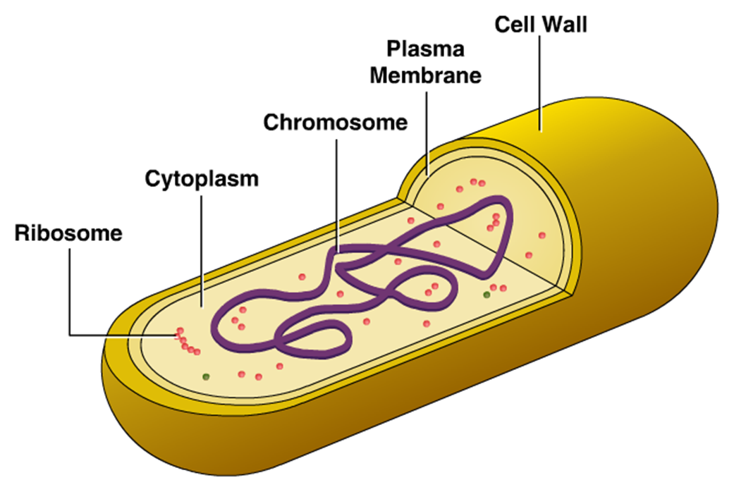
1. A bacterial cell remains surrounded by an outer layer or cell envelope, which consists of two components - a rigid cell wall and beneath it a cytoplasmic membrane or plasma membrane. 2. The cell envelope encloses the protoplasm, made up of the cytoplasm, cytoplasmic inclusions (such as ribosomes, mesosomes, fat globules, inclusion.

Example Image Bacteria Diagram Biology diagrams, Bacteria, Diagram
Easy Bacteria Drawing - Step 2. 2. Draw two long curved lines extending from the cross-section. The lines should be relatively parallel but converge at the end to meet at a point. This is a flagellum, a hair-like structure often compared to a tail. The bacteria uses its flagellum to "swim" or move around.
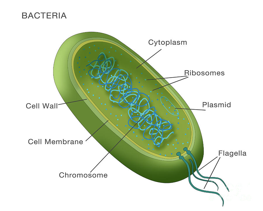
Bacterial Structure Plantlet
Bacteria are diverse, ubiquitous, unicellular, prokaryotic, free-living microorganisms capable of independent reproduction.. Figure 1: Bacteria Cell Diagram.. The small size and simple structure of the bacteria enable them to reproduce rapidly. Theoretically, they can reproduce exponentially until the nutrients are available.
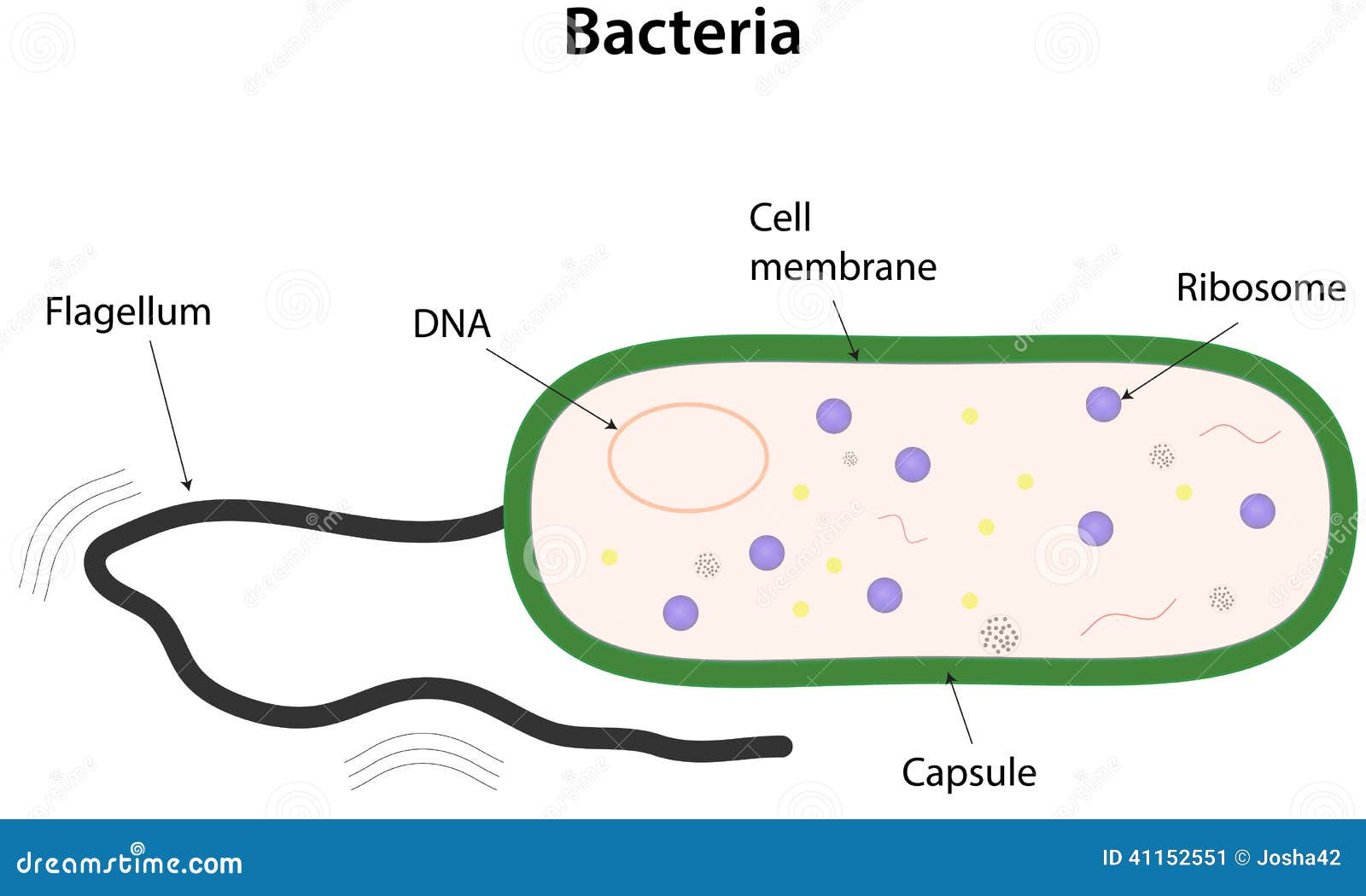
Bacteria Stock Vector Image 41152551
The structure of bacteria is known for its simple body design. Bacteria are single-celled microorganisms with the absence of the nucleus and other c ell organelles; hence, they are classified as prokaryotic organisms. They are also very versatile organisms, surviving in extremely inhospitable conditions. Such organisms are called extremophiles.
Bacteria Diagram Photograph by Monica Schroeder
August 14, 2021. Bacteria are unicellular. Their structure is a very simple type. Bacteria are prokaryotes because they do not have a well-formed nucleus. A typical bacterial cell is structurally very similar to a plant cell. The cell structure of a bacterial cell consists of a complex membrane and membrane-bound protoplast.
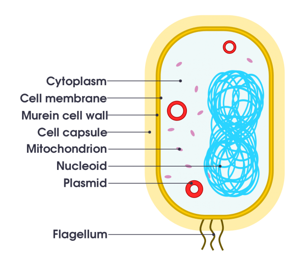
Bacteria Labeled Stock Illustrations 262 Bacteria Labeled Stock Illustrations, Vectors
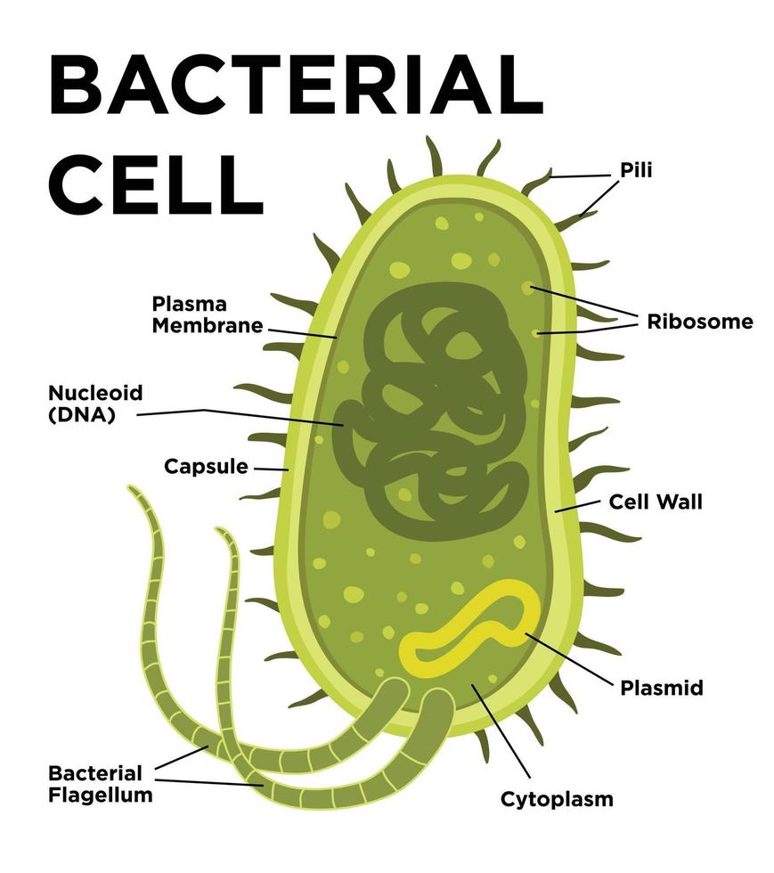
Bacteria diagram can be used to show the structure and shape of the bacterial cell. Here we have used 2D and 3D labelled diagrams and also shape wise classifcation of bacteria.. A simple diagram of a bacterium, labeled in English. It shows the cytoplasm, nucleoid, cell membrane, cell wall, mitochondria, plasmids, flagella, and cell capsule.

Bacterial Cell Diagrams 101 Diagrams
bacteria, any of a group of microscopic single-celled organisms that live in enormous numbers in almost every environment on Earth, from deep-sea vents to deep below Earth's surface to the digestive tracts of humans. Bacteria lack a membrane-bound nucleus and other internal structures and are therefore ranked among the unicellular life-forms.

sdagar1 Year 12 Human Biology Page 2
Cell size. Typical prokaryotic cells range from 0.1 to 5.0 micrometers (μm) in diameter and are significantly smaller than eukaryotic cells, which usually have diameters ranging from 10 to 100 μm. The figure below shows the sizes of prokaryotic, bacterial, and eukaryotic, plant and animal, cells as well as other molecules and organisms on a.
Labelled Diagram Of Bacteria
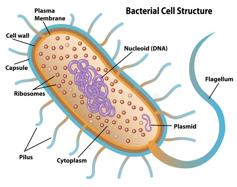
Bacteria - Definition, Structure, Diagram,. The structure of bacteria is relatively simple, yet it allows them to carry out essential functions for their survival. A bacterial cell consists of: Cell Envelope. Cell Envelope is known as glycocalyx which is made up of mucopolysaccharides. Glycocalyx is known as a capsule if it is thick and tough.
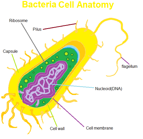
Bacterial cell anatomy in flat style. Vector modern illustration. Labeling structures on a
Size of Bacteria. Bacteria are single-celled organisms. This means that each bacterium is made up of only one cell. This is very different from humans. Our bodies are made up of trillions of cells . Bacteria are much smaller than human cells. Bacterial cells are between about 1 and 10 μm long.
Bacteria Diagram Visual Diagram
Bacterial morphology diagram Types of Bacteria. The cell wall also makes Gram staining possible. Gram staining is a method of staining bacteria involving crystal violet dye, iodine, and the counterstain safranin.. Prokaryote - An organism that has a simple prokaryotic cell; bacteria and archaea are prokaryotes.
